Anthropomorphic Phantoms For Nuclear Medicine
Radiation Equivalent And Anatomically Correct Realistic Test Subjects
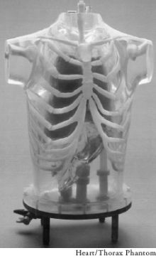
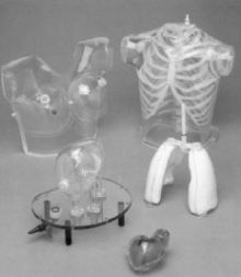
The Heart/Thorax Phantom is ideal for evaluation of detectability, extent and severity of myocardial infarcts in patients.This Phantom also provides valid assessment of mammoscintigraphy techniques.The Striatal Phantom optimizes quantitative imaging in patients, using PET or SPECT.
Myocardial perfusion SPECT is a widely-used, non-invasive method for the diagnosis and management of patients with coronary disease. However, non-uniform photon attenuation, Compton scatter, limited and depth-dependent spatial resolution, as well as image noise, limit the ability of SPECT to obtain images that reliably represent the true tracer distribution.
The non-uniform attenuation of the thorax is the most significant factor limiting the diagnostic efficacy of myocardial SPECT. The currently used attenuation, scatter and resolution correction methods are suboptimal, since they do not provide improvement in the 25% false-negative findings in a group of about 100 patients with luminal diameter stenoses of at least 50%.
Furthermore, the ability to detect multi vessel disease was 70% without and 47% with corrections. This finding implies that myocardial SPECT can seriously underestimate the extent of disease in high risk patients. On the other hand, the false-positive findings in the group with a low probability of coronary disease were reduced from 14% without corrections to 3% with corrections. Obviously, further improvements in both hardware and software for myocardial SPECT are necessary before this important diagnostic technique can realize its full potential. These improvements must be carefully evaluated on realistic, anthropomorphic phantoms to improve results in clinical practice.
FEATURES
- Receptor Quantification As A Function Of Uptake Ratio
- Partial Volume Effects
- Scatter And Attenuation-Correction Schemes
- Threshold for Changes in Uptake
- Comparison Of Different Acquisition Modes, e.g. 2-D Vs 3-D Pet
- Design Of Different Reconstruction Strategies
- Testing And Validation Of Image Registration Techniques
- Design Of Imaging Protocol For Patients
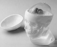
The Head - The Head Phantom is based upon a standard head with a calvarial cut to insert or remove the brain shell easily. The nasal cavity and maxillary sinuses are filled with foam with a mass density of 0.23 g/cc.
Brain Shell - The brain shell has five compartments which can be filled separately: left and right nucleus caudate, left and right putamen, and the remainder of the brain. This allows different nucleus caudate to putamen ratios as well as different striatal to background ratios to be obtained; this also permits differences between left and right striatal activity to be examined.
Livermore Organs -- Organs ( Inert, Hole | Matrix Solid,Active | Inert, 0Hollow )
Lungs - N/A | 5220-RS503A | N/A
Lymph Nodes (set of 3) - N/A | 5220-RS506A | 5220-RS506S
Liver & Liver Envelope - 5220-RS507H | 5220-RS508A (Liver only) | 5220-RS519S (Liver only)
Abdominal Contents* - 5220-RS510H | 5220-RS511A | N/A (*Replaces Liver and Liver Envelope)
Fission-Product Organs -- Organs ( Inert, Hole | Matrix Solid,Active | Inert, 0Hollow )
Lungs - 5220-RS502H | 5220-RS503A | N/A
Lymph Nodes (set of 3) - N/A | 5220-RS506A | 5220-RS506S
Thyroid - N/A | 5220-RS543A | 5220-RS543S
Liver - 5220-RS507H | 5220-RS508A | 5220-RS519S
Kidneys (pair) - 5220-RS530 | H5220-RS531A | 5220-RS532S
Stomach - 5220-RS533H | 5220-RS534A | 5220-RS535S
Pancreas - 5220-RS536H | 5220-RS537A | 5220-RS538S
Spleen - 5220-RS539H | 5220-RS540A | 5220-RS541S
Small Intestine - 5220-RS544H | N/A | N/A
Large Intestine - 5220-RS546H | N/A | N/A


