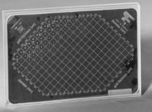
CDMAM 3.4 - CD Phantom for Mammography
In mammography it’s essential that objects with very small contrast and diameter can be distinguished. Therefore the quality of the technical aspects of the mammography equipment should be monitored at regular time-intervals. Usually this is performed by measurement of the physical parameters of the X-ray equipment, screen-film combination, developing process and observation conditions. The main item in quality control however should be the assurance of information transfer from the tissue under examination to the radiologist. Therefore the information content of an image should be monitored.
The CDMAM 3.4 is specially developed to facilitate the evaluation of mammography systems, i.e. detecting very low contrast and very small details. With the phantom the "threshold contrast" as a function of the object diameter can be determined and plotted in a Contrast-Detail curve.
Construction
The CDMAM phantom consists of an aluminum base with gold discs of various thickness and diameter. The aluminum base is attached to a plexiglass (PMMA) cover. Under normal mammography-radiation conditions (Mo anode,30 mm Mo filter, 28 kV) the aluminum base and plexiglass cover together has a equivalent plexiglass thickness of 10 mm. The phantom is delivered with 4 PMMA plates of each 10 mm thickness. Every plate has an engraved marker with lead inlet for identification.
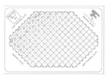
The gold discs are arranged in a matrix of 16 rowsby 16 columns. Within a row the disc diameter is constant, with (partly) logarithmic increasing thickness (see specifications) and within a column the thickness of the discs is constant and the diameter increases logarithmic.
Each square contains two identical discs (same thickness, same diameter), one in the center and one in a randomly chosen corner. Easily recognizable patterns have been avoided.The total matrix is rotated by 45 degrees and the corners of the matrix are skipped. This is done for 2 reasons, getting a better focus on the interesting part (low contrast, small diameter) and making the recognition of the patterns more difficult.
Measurements
To make an X-ray image, the CDMAM phantom (in combination with one or more plexiglass plates) should be positioned on the bucky with the smallest disc diameters at the thorax side. After the film has been processed, the density of the film should be checked. In a series of CD-images, all the images should have approximately the same densities in a reference position on the film.
After this preprocessing, the following measurements are possible
- Comparison of image quality with various film-screen combinations
- Optimization of digital mammography systems
- Determination of the optimum background density, by variation of this density
- Determination of the optimum exposure technique, e.g. by variation of the tube potential
- Comparison of image quality at various thickness, by variation of the amount of plexiglass at a fixed density
Specifications
Base
- Material Aluminum (99,5%)
- Thickness 0.5 mm
- Size 180 x 240 mm (mammography film size)
Gold discs
- Material Gold (99.9999%)
- Thickness 0.03 µm, .. 2.00 µm (16 exponential steps) - contrast range 0,5 - 30% under standard conditions *
- Diameter 0.06..2.0 mm (16 exponential steps)
Cover
- Material - PMMA
- Thickness - together with base, under standard conditions 10 mm plexiglass equivalent
- Size - 180 x 240 mm
- Grid - silkscreen printed with X-ray contrasting paint
Plexiglas plates (4)
- Material - PMMA
- Thickness - 10 mm
- Size - 180 x 240 mm
- Markers - identification markerswith lead inlet
Every phantom is delivered with a reference photo,made under the following standard conditions*:
Tube - Mo anode with 30 mm Mofilter
Potential - 28 kV
Focal spot - large (IEC-grade 0.3)
Grid - present
Exposure time - resulting in D .1.2 O.D. + base and fog
Compression plate - present
The CDMAM 3.4 phantom is a result of the project: "Quality Assurance in Mammography, Department of Radiology, University Medical Centre Nijmegen, (St.Radboud), the Netherlands." By M.A.O. Thijssen, Ph.D., K.R. Bijkerk, M.Sc. and J.M.Lindeyer, B.Sc
0695-0120 CD Phantom for Mammography (CDMAM 3.4)
Works in combination with CDMAM Analyser


